


Review of the application of the open-source software CilOCT for semi-automatic segmentation and analysis of the ciliary muscle in OCT images
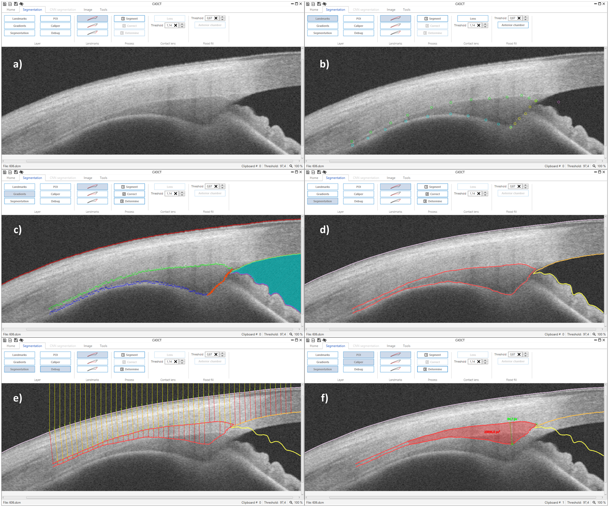
Presbyopia and myopia research shows a growing interest in ciliary muscle biometry using optical coherence tomography (OCT). Until now, segmentation of the ciliary muscle is often performed manually using either custom-developed programs or image processing software. Here we present a novel software for semi-automatic segmentation of the ciliary muscle. It provides direct import of OCT images in DICOM format, a standardized procedure for segmentation, image distortion correction, the export of anatomical ciliary muscle landmarks, like ciliary muscle apex and scleral spur, as well as a continuous thickness profile of the ciliary muscle as a novel way of analysis. All processing steps are stored as XML files, fostering documentation, and reproducibility of research through the possibility of replicating the analysis. Additionally, CilOCT supports batch processing for the automated analysis of large numbers of images and the respective data export to tabulated text files based on the stored XML files. CilOCT was successfully applied in several studies and their results will be summarized in this paper.
